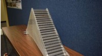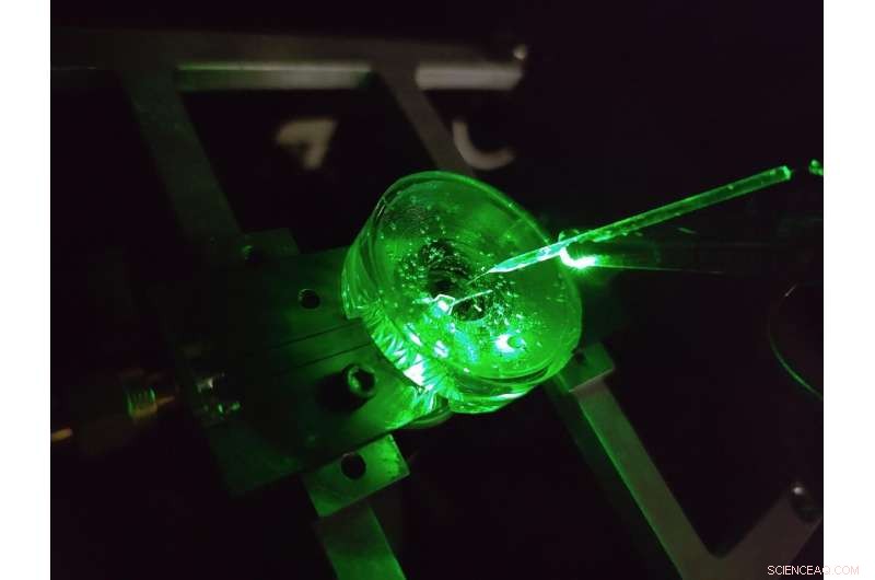
Un prototipo di un microscopio per imaging a voltaggio del diamante costruito dai fisici dell'Università di Melbourne. Un minuscolo elettrodo è sospeso sopra il chip diamantato per testare le prestazioni del dispositivo. Un laser verde illuminato dal basso fornisce eccitazione fluorescente al chip. Credito:Autore fornito, Università di Melbourne
Il cervello è probabilmente una delle strutture più complesse dell'universo conosciuto.
I continui progressi nella nostra comprensione del cervello e nella nostra capacità di trattare efficacemente una serie di malattie neurologiche si basano sull'analisi dei microcircuiti neurali del cervello con dettagli sempre maggiori.
Una classe di metodi per lo studio dei circuiti neurali è chiamata imaging di tensione. Queste tecniche ci consentono di vedere la tensione generata dai neuroni di attivazione del nostro cervello, raccontandoci come le reti di neuroni si sviluppano, funzionano e cambiano nel tempo.
Oggi, l'imaging della tensione di neuroni in coltura viene eseguito utilizzando densi array di elettrodi su cui le cellule vengono coltivate (o coltivate) o applicando coloranti che emettono luce che rispondono otticamente alle variazioni di tensione sulla superficie della cellula.
Ma il livello di dettaglio che possiamo vedere usando queste tecniche è limitato.
Gli elettrodi più piccoli non sono in grado di distinguere in modo affidabile i singoli neuroni, di circa 20 milionesimi di metro, per non parlare della fitta rete di connessioni su nanoscala che si forma tra di loro, e da oltre due decenni non sono stati fatti progressi tecnologici significativi in quest'area.
Inoltre, ogni elettrodo richiede la propria connessione cablata e amplificatore, ponendo notevoli limitazioni al numero di elettrodi che possono essere misurati contemporaneamente.
I coloranti possono superare queste limitazioni immaginando la tensione in modalità wireless come luce:ciò significa che la complessa elettronica può essere situata lontano dalle celle all'interno di una fotocamera.
Il risultato è un'alta risoluzione su vaste aree, in grado di distinguere ogni singolo neurone in una grande rete. Ma anche qui ci sono dei limiti, le risposte di tensione dei coloranti all'avanguardia sono lente e instabili.
La nostra recente ricerca pubblicata su Nature Photonics , esplora un nuovo tipo di piattaforma di imaging a voltaggio ad alta velocità, alta risoluzione e scalabile creata con l'obiettivo di superare queste limitazioni:un microscopio a voltaggio diamantato.
Sviluppato da un team di fisici dell'Università di Melbourne e dell'Università RMIT, il dispositivo utilizza un sensore a base di diamante che converte i segnali di tensione sulla sua superficie direttamente in segnali ottici, ciò significa che possiamo vedere l'attività elettrica mentre si verifica.
The conversion uses the properties of an atom-scale defect in the diamond's crystal structure known as the nitrogen-vacancy (NV).
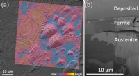
NV defects can be engineered by bombarding the diamond with a nitrogen ion beam using a special type of particle accelerator. The fabrication of the sensor begins with using this process to create a high-density, ultra-thin layer of NV defects close to the diamond's surface.
You can think of each NV defect as a bucket that holds up to two electrons. When this bucket is empty, the NV defect is dark. With one electron, the NV defect emits orange light when illuminated by a laser—this property is known as fluorescence. With two electrons, the color of the fluorescence becomes red.
A previously discovered property of NV defects is that the number of electrons they hold—and the resulting fluorescence—can be controlled with a voltage. Unlike dyes, the voltage response of an NV defect is very fast and stable.
Our research aims to overcome the challenge of making this effect sensitive enough to image neuronal activity.
On the diamond's surface, the crystal structure ends with a layer one atom thick, made up of hydrogen and oxygen atoms. The NV defects closest to the surface are the most sensitive to changes in voltage outside the diamond, but they are also highly sensitive to the atomic makeup of the surface layer.
Too much hydrogen and the NVs are so dark that the optical signals we are looking for cannot be seen. Too little hydrogen and the NVs are so bright that the small signals we are after are completely washed out.
So, there's a "Goldilocks' zone" for voltage imaging, where the surface has just the right amount of hydrogen.
To reach this zone, our team developed an electrochemical method for removing hydrogen in a controlled way. By doing this, we've managed to achieve voltage sensitivities two orders of magnitude better than what has been previously reported.
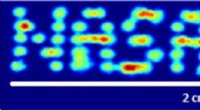
We tested our sensor in salty water using a microscopic wire 10-times thinner than a human hair. By applying a current, the wire can produce a small cloud of charge in the water above the diamond. The formation and subsequent diffusion of this charge cloud produces small voltages at the diamond surface.
By capturing these voltages through a high-speed recording of the NV fluorescence, we can determine the speed, sensitivity and resolution of our diamond imaging chip.
We were able to further boost sensitivity by patterning the diamond's surface into 'nanopillars'—conical structures with the NV centers embedded in their tips. These pillars funnel the light emitted by the NVs towards the camera, dramatically increasing the amount of signal we can collect.
With the development of the diamond voltage imaging microscope for detecting neuronal activity, the next step is the recording of activity from cultured neurons in vitro—these are experiments on cells grown outside their normal biological context, otherwise known as test-tube or petri-dish experiments.
What differentiates this technology from existing state-of-the-art in vitro techniques is the combination of high spatial resolution (on the order of a millionth of a meter or less), large spatial scale (a few millimeters in each direction—which for a network of neurons in mammals is quite vast), and complete stability over time.
No other existing system can simultaneously offer these three qualities, and it's this combination that will allow our made-in-Melbourne technology to make a valuable contribution to the work of neuroscientists and neuropharmacologists globally.
Our system will aid these researchers in pursuing both fundamental knowledge and the next generation of treatments for neurological and neurodegenerative diseases. + Esplora ulteriormente