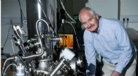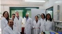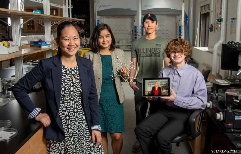
Pinshane Huang, all'estrema sinistra, si unisce a Priti Kharel, il secondo da sinistra, Justin Bae e Patrick Carmichael, all'estrema destra, al Materials Research Laboratory. Nella foto sul tablet c'è Blanka Janicek. Credito:Heather Coit/The Grainger College of Engineering
Uno sforzo di ricerca dell'Università dell'Illinois Urbana-Champaign guidato da Pinshane Huang sta accelerando le tecniche di imaging per visualizzare chiaramente le strutture di piccole molecole, un processo una volta ritenuto impossibile. La loro scoperta libera un potenziale infinito nel miglioramento delle applicazioni della vita quotidiana, dalla plastica ai prodotti farmaceutici.
Il professore associato del Dipartimento di scienza dei materiali e ingegneria ha collaborato con gli autori principali Blanka Janicek, un'allieva del '21 e post-dottorato presso il Lawrence Berkeley National Laboratory a Berkeley, in California, e Priti Kharel, una studentessa laureata del Dipartimento di Chimica, per dimostrare la metodologia che consente ai ricercatori di visualizzare piccole strutture molecolari e accelerare le attuali tecniche di imaging.
Altri coautori includono lo studente laureato Sang hyun Bae e gli studenti universitari Patrick Carmichael e Amanda Loutris. La loro ricerca peer-reviewed è stata recentemente pubblicata in Nano Letters .
Gli sforzi del team espongono la struttura atomica della molecola, consentendo ai ricercatori di capire come reagisce, apprendere i suoi processi chimici e vedere come sintetizzare i suoi composti chimici.
"La struttura di una molecola è così fondamentale per la sua funzione", ha detto Huang. "Quello che abbiamo fatto nel nostro lavoro è rendere possibile vedere direttamente quella struttura."
La capacità di vedere la struttura di una piccola molecola è vitale. Kharel condivide quanto sia vitale citando l'esempio di un farmaco noto come talidomide.
Scoperta negli anni '60, la talidomide veniva prescritta alle donne in gravidanza per curare la nausea mattutina e in seguito si scoprì che causava gravi difetti alla nascita o, in alcuni casi, anche la morte.
Che cosa è andato storto? Il farmaco aveva strutture molecolari miste, una responsabile del trattamento della nausea mattutina e l'altra che purtroppo causava effetti negativi devastanti per il feto.
La necessità di una scienza proattiva, non reattiva ha spinto Huang e i suoi studenti a perseguire questo sforzo di ricerca iniziato originariamente con pura curiosità.
"È fondamentale determinare con precisione le strutture di queste molecole", ha affermato Kharel.
Tipicamente, le strutture molecolari sono determinate con tecniche indirette, un approccio difficile e dispendioso in termini di tempo che utilizza la risonanza magnetica nucleare o la diffrazione dei raggi X. Ancora peggio, i metodi indiretti possono produrre strutture errate che danno agli scienziati la comprensione sbagliata della composizione di una molecola per decenni. L'ambiguità che circonda le strutture delle piccole molecole potrebbe essere eliminata utilizzando metodi di imaging diretto.
In the last decade, Huang has seen significant advancements in cryogenic electron microscopy technology, where biologists freeze the large molecules to capture high-quality images of their structures.
"The question that I had was:What's keeping them from doing that same thing for small molecules?" Huang said. "If we could do that, you might be able to solve the structure (and) figure out how to synthesize a natural compound that a plant or animal makes. This could turn out to be really important, like a great disease-fighter," Huang said.
The challenge is that small molecules are often 100 or even 1,000 times smaller than large molecules, making their structures difficult to detect.
Determined, Huang's students began using existing large molecule methodology as a starting point for developing imaging techniques to make the small molecules' structures appear.
Unlike large molecules, the imaging signals from small molecules are easily overwhelmed by their surroundings. Instead of using ice, which typically serves as a layer of protection from the harsh environment of the electron microscope, the team devised another plan for keeping the small molecules' structures intact.
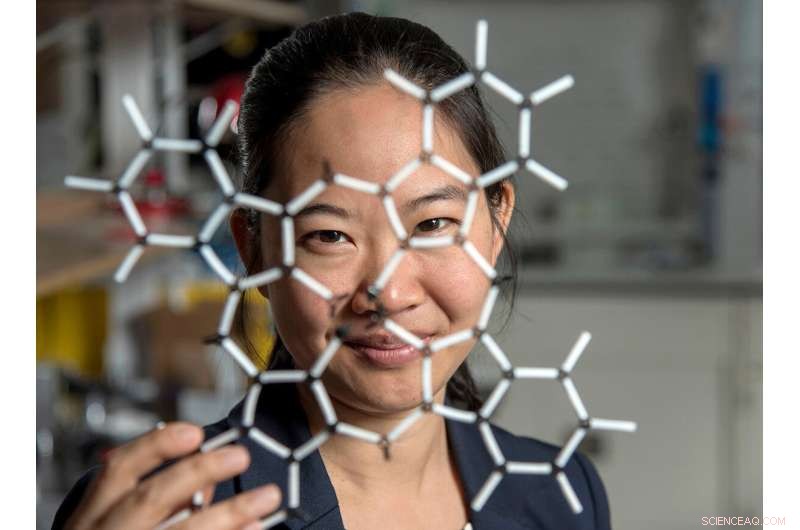
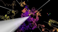
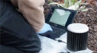
Pinshane Huang and her researchers discovered how to view structures in molecules, which opens a whole new realm of scientific possibilities. Credit:Heather Coit/The Grainger College of Engineering
How can you temper a molecule's environment? By using graphene.
Graphene, a single layer of carbon atoms that form a tight, hexagon-shaped honeycomb lattice, dissipates damaging reactions during imaging.
Stabilizing the small molecule's environment was only one issue the Illinois researchers had to manage. The team also had to limit its use of electrons, as low as one-millionth the number of electrons normally used, to illuminate the molecules.
Low doses of electrons ensure that the molecules are still moving enough for the researchers to capture an image.
"The way I like to think about it is the molecule doesn't like to be bombarded by higher-energy electrons, but we need to do that to be able to see the structure, and graphene helps dissipate some of that charge away from the molecule so that we can actually get a nice image of it," Janicek said.
Unfortunately, once captured, the molecules were nearly invisible in the image.
"When they take a low-dose image, it initially looks like noise or TV static—almost like nothing is there," Huang said.
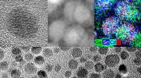
The trick was to isolate the atomic structures from that noise by using a Fourier transform—a mathematical function that breaks down the small molecule's image—to see its spatial frequency.
"We took images of hundreds of thousands of molecules and added them together to build a single, clear image," Kharel said.
This averaging approach allowed the team to create crisp images of the molecules' atoms without damaging the integrity of any individual molecule.
"Month after month, week after week, our resolution improved," Huang said. "And then one day, my students came in and showed me the individual carbon atoms—that's a major achievement. And of course, it comes after all this deep knowledge that they have gained to design an imaging experiment and how to unlock data from what looks like nothing."
This collective discovery is paving the way for many more structural molecule imaging findings.
"There's been this whole field of small molecules that have been left out in the cold, so to speak. We're shining a light on how do we get there as a field? How do we make this thing that for us right now is so hard?" Huang said. "One day it won't be—that's the hope."
The Illinois researchers' efforts are the first big step in turning that dream into reality.
"One day, this will be the way we solve the structure of a small molecule," Huang said. "People will simply throw the molecule in the electron microscope, take a picture and be done."
That dream inspires Huang and her Illinois team to keep the course.
"That's potentially life-changing, and we've made it exist," Huang said. "We haven't yet made it easy, but imaging techniques like this will change so much of science and technology." + Esplora ulteriormente